Waris E (1), Ashammakhi N (2), Lehtimäki M (3),
Tulamo R-M (4),
Kellomäki M (5), Törmälä P (5), Santavirta S (6), Konttinen YT
(1).
1. Institute of Biomedicine/ Anatomy, Biomedicum Helsinki, University
of
Helsinki, Finland;
2. Division of Plastic Surgery, Department of Surgery, Oulu University
Hospital;
3. Department of Rheumatoid Surgery and Orthopedics, Tampere University
Hospital;
4. Department of Clinical Veterinary Sciences, Faculty of Veterinary
Medicine,
University of Helsinki;
5. Institute of Biomaterials, Tampere University
of Technology;
6. Department of Orthopaedics and Traumatology, Helsinki University Central
Hospital.
Silicone interposition arthroplasty is
the most commonly used method for reconstruction of metacarpophalangeal
(MCP) joints. Swanson silicone arthroplasty restores hand function in
appropriately selected patients. However, decreased mobility, recurrent
pain and instability become prevalent with long-term follow-up. Reported
complications include infection, implant dislocation and fracture [1].
The lack of bone stock may cause a formidable challenge in revision arthroplasty.
Modern tissue engineering technology has led to the development of bioreplaceable
scaffolds for reconstruction of small joints of the hand [2]. This experimental
study is undertaken to evaluate biological behavior, bioabsorption and
biocompatibility of bioreplaceable and Swanson silicone interposition
arthroplasties performed in the MCP joints of minipigs using histological,
microradiographic, radiographic and range of joint motion (ROM) analyses.
Materials and methods
Two types of bioabsorbable implants, a cylindrical scaffold
made of poly-L/D-lactide (L,D-monomer ratio of 96/4, P(L/D)LA 96/4) and
a P(L/D)LA 96/4 scaffold combined with a Polyactive® stem, were used in
this study. Raw P(L/D)LA 96/4 (Purac biochem B.V. Gorinchem, Netherlands)
was melt-spun into a 4-ply multifilament, knitted to tubular jersey and
then rolled to a cylindrical scaffold Ø8 x 3.5 mm in size. The stemmed
P(L/D)LA 96/4 implant had an elastomer-like stem made of a copolymer of
polyethylene glycol terephalate (PEGT) and polybutylene terephalate (PBT),
Polyactive®, having a PEGT/PBT proportion of 70/30 (H.C. Implants B.V.,
Netherlands). The silicone Swanson finger joint implant (Wright Medical
Technology, Inc.) of size 00 was appropriate to meet the anatomical requirements.
18 skeletally mature female minipigs were operated. The operation was
performed on both fore hooves under tourniquet control in general anaesthesia.
A longitudinal incision was made over the dorsum of the fifth MCP joint.
After the joint was exposed, the metacarpal head and the base of proximal
phalanx were resected, leaving the collateral ligaments intact. All cartilage
was removed to simulate end-stage rheumatoid arthritis. The joint reconstruction
was achieved with one of three different implants described above: 1)
P(L/D)LA 96/4 scaffold, 2) P(L/D)LA 96/4 scaffold with a Polyactive® stem,
or 3) Swanson silicone implant. Resection without any implantation serves
as the fourth study group.
Throughout the postoperative course the joint is assessed by radiological
and range of motion (ROM) examination. The animals are sacrificed at 12
and 24 weeks as well as at 1 and 3 years to obtain three specimens from
each study group. The bone specimens are fixed in a series of ethanol
immersions of rising concentrations and embedded in methylmetacrylate.
For histological microradiographic and OTC-fluorescence studies, longitudinal
sections are cut with a microtome. The axillary lymph nodes are evacuated,
and also biopsies of the liver and spleen are performed to demonstrate
histologically possible lymphadenopathy and silicone granulomas.
Results and discussion
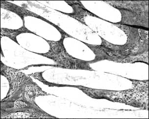
All 18 minipigs have been operated in autumn 2001. Cylindrical
P(L/D)LA 96/4 implant serves as a temporary scaffold for fibrous cell
invasion and have shown good biocompatibility during the follow up of
10 weeks. Fluid accumulation and sinus formation was seen in all radiographs
at 10 and 26 weeks following Polyactive® stem. Swanson silicone implants
were surrounded by fibrous capsule. Silicone wear particles were seen
in the interface tissue at 10 weeks and were migrated to regional lymph
nodes.
The animals are followed up to three years when the P(L/D)LA
96/4 scaffold should be completely absorbed and replaced by the animals
own tissue. The bioreplaceable, biocompatible, P(L/D)LA 96/4 scaffold
provides apparently a resilient, functional MCP arthroplasty allowing
unlimited use of the joint and avoiding long-term complications and restrictions
of current biostable prostheses.
References
[1] Vasenius J et al. Current Orthopaedics 14, 284-289,
2000;
[2] Lehto MUK, Lehtimäki, MY, Paasimaa S, Törmälä, P: PCT Pat. Appl. No.
FI96/00035;
[3] Honkanen P et al. Biodegradable PLDLA scaffold prosthesis in the MCP-arthroplasty
of rheumatoid patients. SOT 2, 23, 111, 2000