Hyperspectral Imaging
Hyperspectral imaging assisted with the ML technique has been applied for functional skin imaging
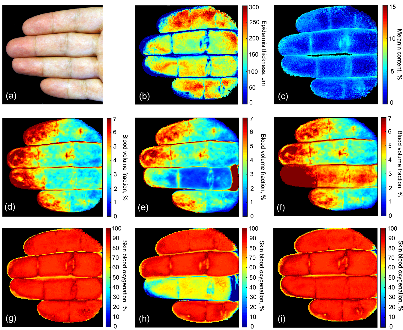
Reconstructed values of the epidermal thickness (b) and melanin content (c) for the Caucasian skin type (a). Retrieved maps of skin blood volume fraction before (d), during (e) and 1 min after (f) the ring finger occlusion. Increase of skin blood volume fraction after the release of the occlusion ring (f) clearly indicated the reactive hyperemia. Corresponding maps of skin blood oxygenation before (g), during (h) and 1 min after (i) the occlusion.
Monitorig of skin complications of Diabetes Mellitus with Polarized Hyperspectral imaging.

The developed system has been used to reveal early changes in skin blood microcirculation and skin structure of patients with diabetes. The dorsal surface of the patients’ feet has been imaged.
It was observed that the diabetic patients had increased skin blood content and, at the same time, the reduced oxygen level in comparison to the control group of healthy volunteers. In addition, the diabetic group has an increased polarization index that is attributed to the changes in skin collagen structure.

The values of blood volume fraction, skin blood oxygenation, and polarization index were found to have statistically significant difference between the control and the diabetic groups.
1. V. Dremin, Z. Marcinkevics, E. Zherebtsov, A. Popov, A. Grabovskis, H. Kronberga, K. Geldnere, A. Doronin, I. Meglinski, A. Bykov, "Skin complications of diabetes mellitus revealed by polarized hyperspectral imaging and machine learning", IEEE Trans. Med. Imaging, 40(4), 1207-1216 (2021).
2. V. Dremin, E. Zherebtsov, A. Bykov, A. Popov, A. Doronin, and I. Meglinski, "Influence of blood pulsation on diagnostic volume in pulse oximetry and photoplethysmography measurements," Appl. Opt. 58, 9398-9405 (2019).
3. E. Zherebtsov, V. Dremin, A. Popov, A. Doronin, D. Kurakina, M. Kirillin, I. Meglinski, A. Bykov, “Hyperspectral imaging of human skin aided by artificial neural networks”, Biomed. Opt. Express, 10(7), 3545-3559 (2019). (top downloads Jul 2019, noteworthy research highlight 2021).
4. A. Popov, A. Bykov, I. Meglinski, “Influence of probe pressure on diffuse reflectance spectra of human skin measured in vivo” J. Biomed. Opt., 22(11), 110504 (2017). (cover page article).
5. J. Spigulis, I. Oshina, A. Berzina, A. Bykov, “Smartphone snapshot mapping of skin chromophores under triple-wavelength laser illumination”, J. Biomed. Opt., 22(9), 091508 (2017).